
Quick Takeaways
- Reperfusion restores blood flow but unleashes reactive oxygen species (ROS) that damage cells.
- Damaged cells release DAMPs that activate Toll‑like receptors (TLRs) and a cytokine storm.
- Persistent inflammation can break self‑tolerance, leading to diseases such as SLE and RA.
- Clinical studies link heart attacks, stroke, and organ transplantation to later autoimmune phenomena.
- Targeting ROS, DAMP‑TLR signaling, or specific cytokines shows promise in preventing autoimmunity after reperfusion.
What Is Reperfusion Injury?
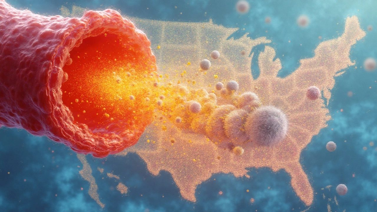
Reperfusion Injury is a type of tissue damage that occurs when blood flow returns to an area that was previously ischemic. The sudden oxygen influx triggers bursts of reactive oxygen species (ROS), endothelial dysfunction, and a rapid inflammatory response.
Clinically, you see it after heart attacks, strokes, or organ transplants. Troponin spikes, edema, and microvascular blockage are hallmark signs.
Key Players in the Injury Cascade
Several molecular actors turn a life‑saving reperfusion into a double‑edged sword.
- Reactive Oxygen Species are highly reactive molecules (e.g., superoxide, hydrogen peroxide) that oxidize lipids, proteins, and DNA.
- Damage‑Associated Molecular Patterns (DAMPs) are intracellular fragments released by dying cells that act as danger signals.
- Toll‑like Receptors (TLRs) on immune cells recognize DAMPs, igniting NF‑κB signaling and cytokine production.
- Cytokine Storm refers to the massive release of pro‑inflammatory cytokines (IL‑1β, IL‑6, TNF‑α) that amplify tissue injury.
From Inflammation to Autoimmunity
Why does a burst of inflammation sometimes turn the immune system against itself? The answer lies in three interconnected mechanisms.
- Epitope Spreading: ROS‑modified proteins generate new antigenic epitopes that were never seen by the immune system before.
- Molecular Mimicry: Some altered self‑peptides closely resemble microbial antigens, causing cross‑reactive T‑cells to attack host tissue.
- Regulatory T‑cell Impairment: The oxidative environment hampers Treg function, reducing the brakes on auto‑reactive lymphocytes.
When these processes persist, they can seed long‑lasting autoimmune disorders.
Autoimmune Disorders Linked to Reperfusion Injury
Evidence from both animal models and human cohorts ties reperfusion to specific autoimmune diseases.
- Systemic Lupus Erythematosus (SLE) patients show higher anti‑oxidized‑LDL antibodies after myocardial infarction, suggesting ROS‑driven autoantigen formation.
- Rheumatoid Arthritis flares have been reported following limb revascularization surgery, correlating with elevated serum HMGB1 (a DAMP).
- Experimental autoimmune encephalomyelitis (EAE) models demonstrate that cerebral reperfusion after stroke accelerates demyelination via TLR‑4 activation.
Clinical Evidence and Biomarkers
Large‑scale registry analyses (e.g., the INTERHEART and TRANSPLANT cohorts) reveal a 1.8‑fold increase in new‑onset autoimmune diagnoses within five years after acute reperfusion events.
Key biomarkers that flag a transition from injury to autoimmunity include:
- Serum levels of high‑mobility group box 1 (HMGB1) - a prototypical DAMP.
- Elevated anti‑oxidized lipid antibodies measured by ELISA.
- Persistent rise in IL‑17 and IFN‑γ, indicating Th17 skewing.
Therapeutic Strategies: Breaking the Cycle
Intervening early can prevent the autoimmune sequelae of reperfusion injury.
| Aspect | Reperfusion Injury | Autoimmune Disorder |
|---|---|---|
| Primary Trigger | Sudden oxygen influx after ischemia | Loss of self‑tolerance |
| Key Mediators | ROS, DAMPs, TLR activation | Auto‑antibodies, autoreactive T‑cells |
| Typical Clinical Context | Myocardial infarction, stroke, transplantation | Systemic lupus, rheumatoid arthritis, multiple sclerosis |
Current therapeutic avenues focus on three fronts:
- Antioxidants: N‑acetylcysteine or mito‑TEMPO administered during reperfusion reduce ROS‑mediated epitope formation.
- TLR Antagonists: Oligonucleotide‑based TLR‑7/9 inhibitors dampen DAMP signaling, limiting cytokine storms.
- Targeted Immunomodulation: Low‑dose IL‑6 blockers (e.g., tocilizumab) given post‑reperfusion curb Th17 expansion without broad immunosuppression.
Combination regimens-antioxidant plus TLR blockade-have shown the best outcomes in phase‑II trials for post‑myocardial infarction patients.
Related Concepts and Future Directions
Understanding the bridge between reperfusion injury and autoimmunity opens doors to several adjacent topics:
- Ischemia‑Reperfusion (I/R) Injury in organ transplantation-how pre‑conditioning may lower long‑term graft‑related autoimmunity.
- Neutrophil Extracellular Traps (NETs) as another source of autoantigens after reperfusion.
- Complement System Activation and its role in amplifying tissue damage and autoantibody production.
- Epigenetic Reprogramming of immune cells in the oxidative microenvironment, which may lock in autoreactive phenotypes.
Researchers are now probing nanocarrier‑delivered ROS scavengers that release directly at the reperfused vasculature, hoping to neutralize danger signals before they awaken the adaptive immune system.
Practical Take‑Home Points for Clinicians
- Monitor high‑risk patients (post‑MI, stroke, transplant) for early biomarkers like HMGB1 and anti‑oxidized‑LDL antibodies.
- Consider peri‑reperfusion antioxidant therapy, especially in patients with a personal or family history of autoimmunity.
- If autoimmune symptoms emerge, evaluate the timeline-symptom onset within months of reperfusion suggests a causal link.
- Collaborate with rheumatology to tailor immunomodulatory treatment that addresses both injury‑driven inflammation and emerging autoimmunity.
Frequently Asked Questions
Can reperfusion injury cause permanent autoimmune disease?
Not every episode leads to a chronic autoimmune disorder, but persistent oxidative stress and unchecked inflammation can tip the balance toward autoimmunity. Long‑term follow‑up studies show a higher incidence of diseases like SLE in patients who experienced severe reperfusion injury.
What are the most reliable biomarkers to watch after an infarct?
HMGB1, anti‑oxidized‑LDL antibodies, and sustained elevations of IL‑6 or IL‑17 are the best early signals. Serial measurements over the first two weeks can guide whether prophylactic immunomodulation is warranted.
Are antioxidants effective in preventing autoimmunity after reperfusion?
Clinical trials with N‑acetylcysteine and mito‑TEMPO report reduced ROS levels and fewer auto‑antibody spikes, but results vary by dosage and timing. The consensus is that antioxidants are most effective when given right at the moment of reperfusion.
How do Toll‑like receptor inhibitors work in this context?
TLR antagonists block DAMP‑driven NF‑κB activation, curbing the cytokine surge that fuels both tissue damage and auto‑reactive lymphocyte activation. Early‑phase studies show fewer post‑procedural flares when TLR‑7/9 inhibitors are added to standard care.
Should patients with existing autoimmune disease receive special reperfusion protocols?
Yes. Those patients benefit from intensified antioxidant regimens and pre‑emptive low‑dose immunomodulators to prevent disease exacerbation. Tailoring the approach reduces the risk of a sudden flare triggered by the reperfusion insult.

Gurupriya Dutta
September 22, 2025 AT 09:44Especially the part about epitope spreading-like the body’s own cells are being rewritten into foreign invaders. Scary, but also a huge opportunity for early intervention.
Michael Lynch
September 23, 2025 AT 08:45caroline howard
September 23, 2025 AT 09:24Melissa Thompson
September 24, 2025 AT 02:57Rika Nokashi
September 24, 2025 AT 06:06Don Moore
September 24, 2025 AT 16:08Austin Levine
September 25, 2025 AT 03:40Matthew King
September 25, 2025 AT 23:23james landon
September 26, 2025 AT 15:30Jenn Clark
September 26, 2025 AT 20:40L Walker
September 27, 2025 AT 08:37Andrea Swick
September 28, 2025 AT 03:36Amelia Wigton
September 28, 2025 AT 20:33Meredith Poley
September 29, 2025 AT 05:38Mathias Matengu Mabuta
September 29, 2025 AT 13:39Ikenga Uzoamaka
September 29, 2025 AT 14:01Lee Lee
September 30, 2025 AT 12:16John Greenfield
September 30, 2025 AT 15:49Dr. Alistair D.B. Cook
September 30, 2025 AT 20:27